Student Log in
 1267 Cornwall Rd, Unit 300, Oakville, Ontario L6J 7T5
 (289) 271-5718
blog
What Happens During a Vascular Occlusion – Understanding Tissue Ischemia in Dermal Fillers

Feb 23 2026
Reading Time: 6 Minutes
Author:
Vascular occlusion is one of the most feared complications in medical aesthetics. It is not common, but it can progress quickly. For injectors, understanding vascular occlusion is not just about emergency response. It is about understanding what happens inside the tissue in real time. It is about how ischemia develops and why timing matters so much.
When a vascular occlusion occurs, tissue starts a cascade of physiological changes. These changes can move from reversible to irreversible rapidly. In high-risk areas like the lips, this progression is even faster. Understanding the pathophysiology behind vascular occlusion lip filler complications helps injectors recognize subtle signs earlier. They can respond with confidence and protect both patients and professional integrity.
This blog explores what happens inside the tissue during a vascular occlusion. It explains how ischemia progresses. It also shows why advanced anatomical and complication training is essential for anyone performing dermal filler injections.
What Vascular Occlusion Really Means
At its core, vascular occlusion means blood flow through a vessel is blocked. In dermal fillers, this blockage happens in two main ways.
The first is intravascular injection. Filler enters a blood vessel directly and blocks circulation from within. The second is extrinsic compression. Filler placed near a vessel exerts enough pressure to collapse it and restrict blood flow.
In both cases, the result is the same. Oxygenated blood no longer reaches the tissue supplied by that vessel. Without oxygen, cells cannot produce energy. Without energy, cellular function deteriorates rapidly.
This loss of oxygen delivery is known as ischemia.

All images used under license from Canva. © APT Medical Aesthetics, 2026. All rights reserved.
Why Ischemia Is So Dangerous in Dermal Filler Treatments
Ischemia is not simply reduced blood flow. It is a state where tissue suffers injury from lack of oxygen and nutrients. Unlike other tissues, facial tissues have limited reserves.
The lips are highly metabolic and depend on continuous perfusion. When ischemia occurs in lip tissue, cellular injury can begin within minutes.
The danger lies in both the initial occlusion and the biological cascade that follows.
The Immediate Tissue Response to Vascular Occlusion
When blood flow stops, the first change is hypoxia. Oxygen delivery to cells decreases. Cells shift from aerobic to anaerobic metabolism. This process is inefficient and produces lactic acid.
As lactic acid builds up, tissue pH drops. This acidic environment damages cellular enzymes and cell membranes. Waste products are no longer cleared due to poor circulation.
Clinically, this early phase may show as pain, blanching, or subtle color changes. These are the earliest warning signs injectors may observe.

All images used under license from Canva. © APT Medical Aesthetics, 2026. All rights reserved.
Why Pain Often Appears Early
Pain is often one of the first signs of ischemia. Ischemic pain is different from typical injection discomfort. It is intense, persistent, and progressive.
This pain happens because nerve endings are sensitive to hypoxia and metabolic byproducts. As oxygen drops and lactic acid builds, nerves become irritated and signal distress.
In vascular occlusion lip filler cases, escalating pain should never be ignored. It is often the body’s first alarm that tissue is being deprived of blood.

All images used under license from Canva. © APT Medical Aesthetics, 2026. All rights reserved.
The Progression From Hypoxia to Cellular Injury
If ischemia continues, cellular damage speeds up. Mitochondria, which make energy for the cell, start to fail. Without enough ATP, cells cannot keep their membranes intact.
As cell membranes break down, intracellular contents leak out. This triggers inflammation. Swelling increases and further compresses surrounding vessels, making ischemia worse.
This creates a vicious cycle. Ischemia leads to swelling, and swelling leads to more ischemia.
Why Blanching and Color Changes Occur
Blanching is a visible sign of reduced blood flow. When blood stops flowing through capillaries, the skin looks pale or white.
As ischemia gets worse, color changes may evolve. Tissue may look dusky, mottled, or violaceous. These changes show uneven blood flow, venous congestion, and tissue injury.
In vascular occlusion lip filler events, these color changes may follow arterial patterns. Recognizing these patterns requires anatomical knowledge and clinical experience.
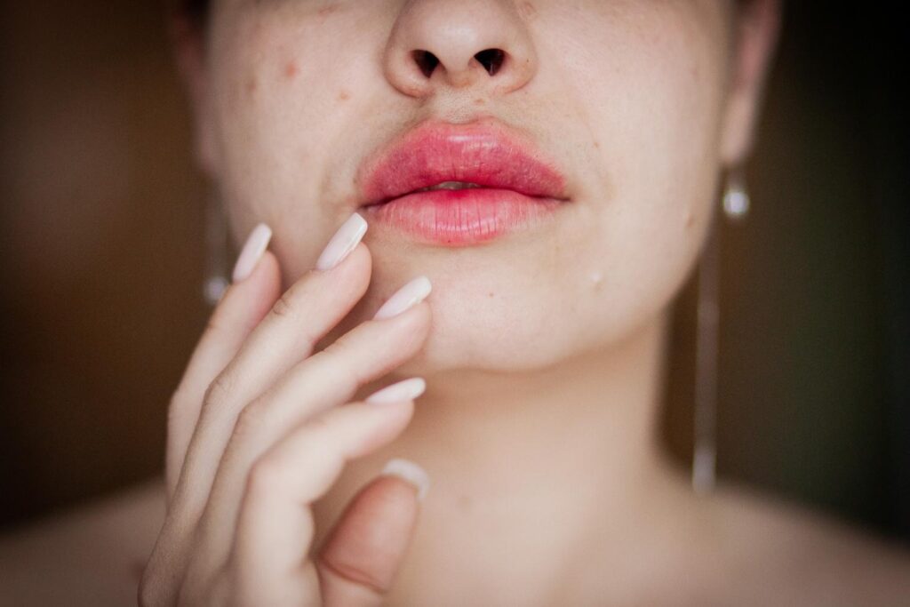
All images used under license from Canva. © APT Medical Aesthetics, 2026. All rights reserved.
Capillary Refill and Perfusion Failure
Capillary refill directly reflects tissue perfusion. In healthy tissue, blood quickly returns to capillaries after pressure is released.
During ischemia, refill is delayed or absent. This means blood flow is not enough to meet tissue needs.
Capillary refill testing gives injectors an objective measure of perfusion. It should be used whenever vascular compromise is suspected.
The Transition from Reversible to Irreversible Injury
One key concept injectors must understand is that ischemic injury follows a timeline.
In the early phase, ischemia may be reversible. Restoring blood flow during this window can allow full recovery.
As ischemia continues, cells cross a threshold. Recovery is no longer possible. Cell death occurs, leading to necrosis. Once necrosis develops, tissue loss is inevitable.
The exact timing varies based on tissue type, blood supply, and patient factors. But facial tissues generally have a narrow window for intervention.
This is why time is the most critical variable in managing vascular occlusion.

All images used under license from Canva. © APT Medical Aesthetics, 2026. All rights reserved.
Why Lip Tissue Is Especially Vulnerable
The lips are supplied by small arterial branches with limited collateral circulation. There are few alternative routes for blood flow if one vessel is blocked.
Lip tissue is also thin and lacks protective fat layers. Swelling in this confined space increases pressure rapidly, making ischemia worse.
These factors make vascular occlusion lip filler complications very urgent and unforgiving.
Inflammation and Secondary Tissue Damage
As ischemia progresses, inflammation gets stronger. Inflammatory mediators attract immune cells, which release enzymes and free radicals.
Inflammation is part of healing, but in ischemia it often makes damage worse. Increased vascular permeability leads to edema, which further compresses vessels.
This secondary damage shows why early intervention is far more effective than managing late-stage complications.

All images used under license from Canva. © APT Medical Aesthetics, 2026. All rights reserved.
Necrosis and Its Consequences
If ischemia is not resolved, tissue necrosis develops. Necrotic tissue loses structural integrity and becomes vulnerable to infection.
In the lips, necrosis can result in:
- Scarring
- Contour deformities
- Loss of tissue
- Functional impairment
- Long-term aesthetic consequences
These outcomes are devastating for patients and emotionally challenging for injectors.
A Clinical Scenario That Illustrates Progression
An injector performs lip filler on a patient. Shortly after, the patient reports increasing pain. The injector sees blanching along the upper lip.
If action is taken immediately, blood flow can be restored and tissue can recover.
If the injector waits, swelling increases, pain worsens, and color changes progress. Hours later, the tissue becomes dusky and cold. Necrosis follows.
The difference between these outcomes lies in understanding and responding to ischemia early.
Why Understanding Pathophysiology Improves Decision Making
Injectors who understand what happens during a vascular occlusion make better decisions. They recognize patterns and act decisively instead of relying on hope.
Understanding tissue ischemia changes complication management from reactive to proactive.

All images used under license from Canva. © APT Medical Aesthetics, 2026. All rights reserved.
Why Complication Training Must Go Beyond Protocols
Emergency protocols are essential, but they are not enough alone. Injectors must understand why they perform each step and how it affects tissue.
APT Injection Training emphasizes this deeper understanding. We teach the mechanisms behind complications, not just the response steps.
This approach builds confidence and reduces hesitation in real-world scenarios.
Tips to be a
Successful Injector!
Free E-Book download